Imaging in Intensive Care Medicine
"Air in the water"—the hydropoint sign
Eleni Soilemezi, Petros Morfesis, Vassiliki Birmpa, et al
Intensive Care Med 2025; 51: 1721–1723
A 30-year-old female patient was admitted in ICU due to community-acquired pneumonia causing bilateral lung infiltrations and severe hypoxemia requiring mechanical ventilation. An episode of rapid desaturation (SpO2 87% despite increased FiO2 to 100%) triggered a new lung ultrasound assessment, which revealed the presence of hydropneumothorax, i.e., both air and fluid were present in the pleural cavity. The typical ultrasonographic finding in hydropneumothorax is called hydropoint and does not share the same features with lung point, the finding establishing the diagnosis of pneumothorax. Instead, the hydropoint sign describes the dynamic change between a pneumothorax pattern and a pattern of pleural effusion during respiration, corresponding to the interface between air and fluid in the pleural cavity. Therefore, a repetitive change from parietal pleural with A-lines only (and no signs of lung tissue such as sliding, B-lines, or lung pulse), to pleural effusion (Fig. 1a, b, ESM video), was demonstrated. Insertion of a chest drain restored the patient’s oxygen saturation.

A Demonstration of parietal pleura and pleural effusion at end-expiration. B Without moving the transducer, A-lines appear at endinspiration at the exact site where pleural fluid was previously seen. It should be noted that A-lines are not accompanied by other signs of lung tissue, such as B-line(s) or lung sliding. The alternating pattern from pleural effusion to parietal pleura with A-lines during different phases of the respiratory cycle, constitutes the hydropoint sign, which denotes the presence of hydropneumothorax. C The scanning site where the hydropoint sign was identified, located posteriorly to the midaxillary line. D The chest X-ray performed immediately after the ultrasound finding of hydropneumothorax. E, F A small lung point is identified only at the apex of the lung immediately after insertion of a midaxillary chest drain, corresponding to a remaining small apical pneumothorax. The lung point clearly separates the presence of a small remaining pneumothorax (demonstrated as visible parietal pleura with reverberating A-lines only), from lung tissue (visceral pleura with B-lines)
Conclusively, the hydropoint (Fig. 2) sign is an ultrasonographic sign of dynamic nature, distinctly different from lung point. The specific clinical setting, where the sign is recognized, determines its significance and urgency to treat.
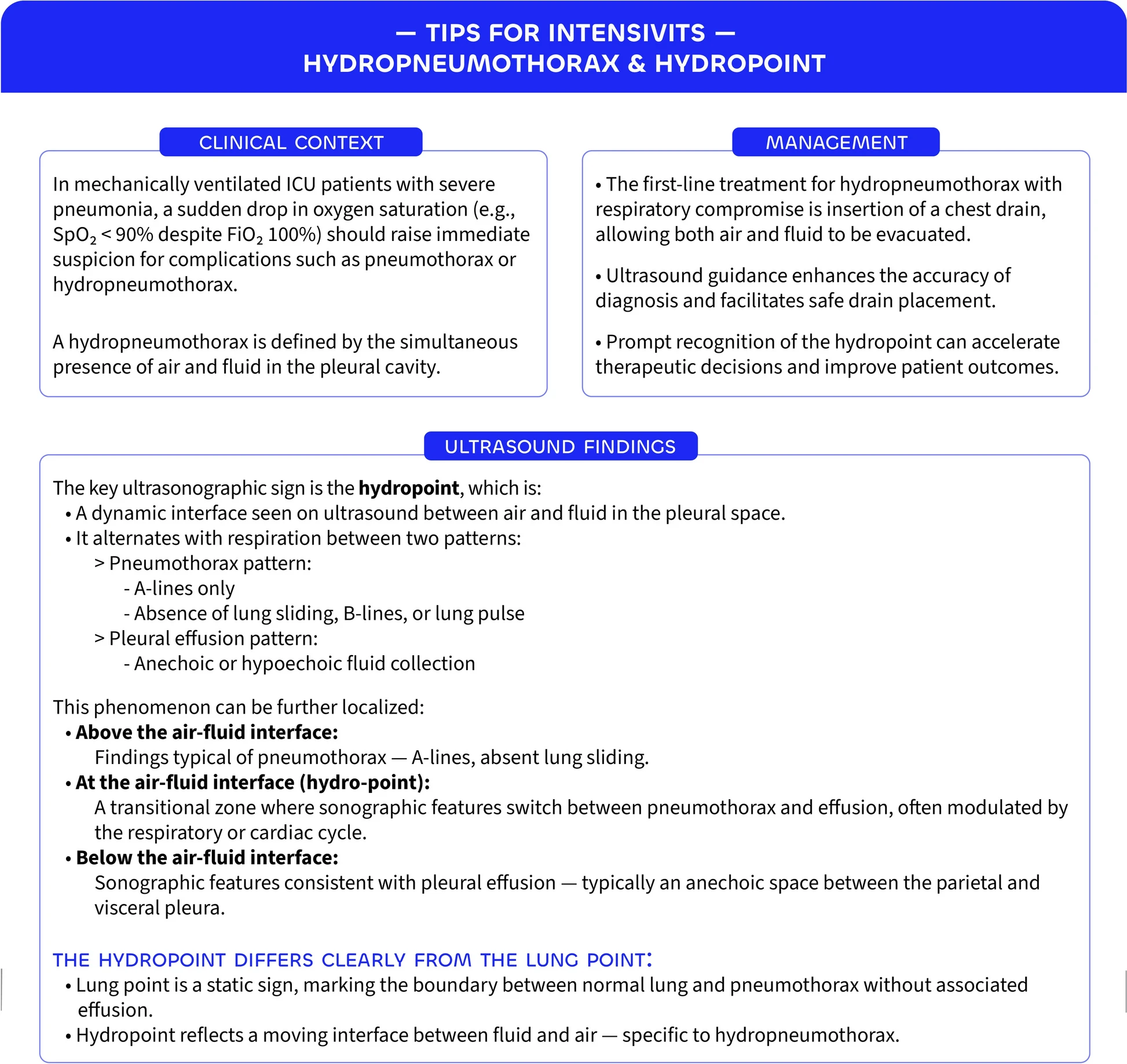
Hydropneumothorax and hydropoint – Tips for intensivists