Imaging in Intensive Care Medicine
Hypervirulent Klebsiella pneumoniae: watch the eyes
Yan Wang, He Zhou, Meiling Ding, et al
Intensive Care Med (2025). https://doi.org/10.1007/s00134-025-08109-3
Imaging studies (Fig. 1, Panels A–E) revealed a solitary lung abscess, a large hepatic abscess, and endophthalmitis characterized by flocculent vitreous opacities. Liver puncture drainage was performed. Microbiological cultures (Panel F) from blood, liver aspirate, and vitreous fluid were all positive for hypervirulent Klebsiella pneumoniae(hvKP). Echocardiography revealed no valvular vegetations. Systemic antibiotic therapy was initiated with intravenous meropenem, followed by piperacillin-tazobactam, along with an intravitreal injection of ceftazidime (0.1 mL/2 mg) in the left eye.
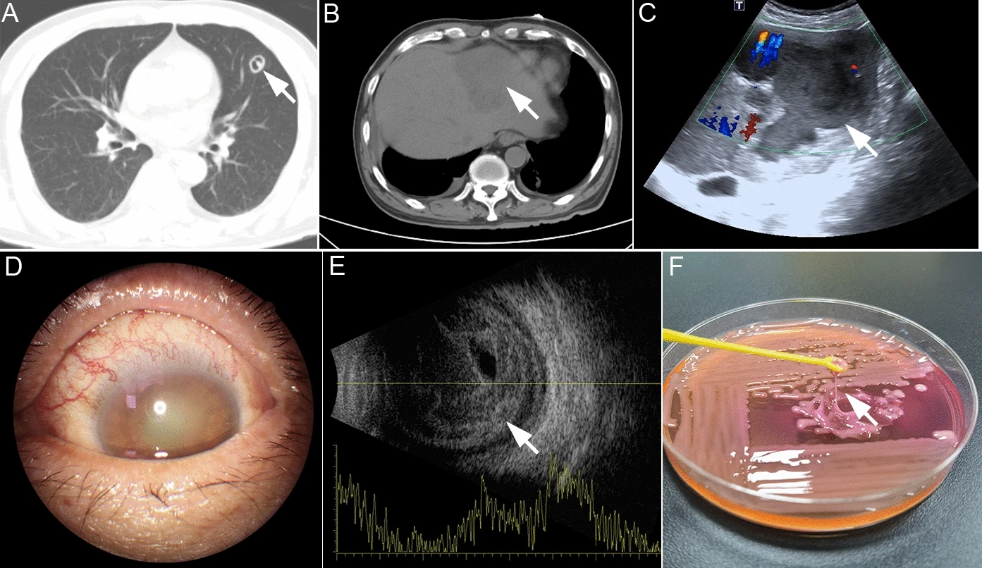
hvKP is a life-threatening pathogen that requires prompt clinical recognition. It typically manifests as a community-acquired pyogenic liver abscess complicated by bacteremia [1, 2]. Ocular involvement is a common complication of disseminated hvKP infection [1], and visual symptoms may progress rapidly to blindness despite aggressive systemic and intravitreal antibiotic therapy. Such metastatic infections are often associated with host risk factors such as diabetes mellitus. Although the string test can provide presumptive evidence of hypermucoviscosity, it is not definitive. Molecular confirmation—such as detection of virulence genes rmpA or magA—is recommended for definitive hypervirulence typing [2]. In this case, virulence gene analysis of the liver aspirate confirmed the presence of iucA and rmpA2.