IMAGES IN CLINICAL MEDICINE
Aspiration of a Chicken Bone
Huey Ming Seah, Marcela Mautone
N Engl J Med 2018; 378:e25
DOI: 10.1056/NEJMicm1713423
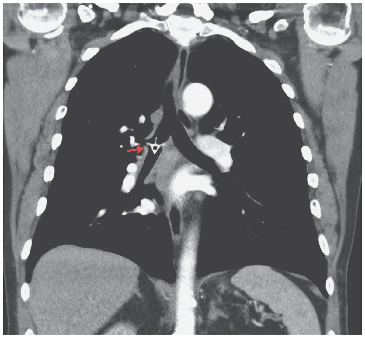
A 78-year-old man presented to the emergency department with stridor, shortness of breath, and fever (38.1°C). Five days earlier, he had come to the emergency department with the sensation of having a foreign body in his throat, approximately an hour after a choking episode that had occurred while he was eating chicken. At that time, the physical examination and plain radiographs of the neck and chest were unremarkable. The foreign body was presumed to have been dislodged, and the patient was discharged home. At the current presentation, computed tomography revealed a high-attenuation structure in the shape of a chicken vertebra in the right mainstem bronchus (arrow). There was minor atelectasis in the right lower lobe without evidence of lobar collapse. Foreign bodies more commonly become lodged in the right side of the bronchial tree than in the left because of its more vertical path and wider lumen. Delayed presentation because of minor symptoms can lead to inflammation and infection. Bronchoscopy was performed, and the chicken bone was successfully removed. The patient recovered well after the procedure and was discharged home on day 3.
78岁男性患者因喘鸣、呼吸困难及发热(38.1°C)到急诊就诊。5天前,患者在吃鸡过程中出现呛咳,约一小时后因咽部异物感到急诊就诊。当时的体格检查及颈胸部X线未发现明显异常。医生考虑异物已经移位,因此允许患者回家。此次就诊CT显示右主支气管内有一高密度结构,形状极似鸡脊椎骨(箭头)。右下叶轻度肺不张,没有肺叶塌陷的表现。由于右主支气管较左主支气管角度更为垂直且管腔更粗,因此异物通常卡在右侧。由于临床症状较轻导致患者就诊延误,可引起炎症和感染。患者接受支气管镜检查,成功取出鸡骨。随后患者恢复良好,第三天出院回家。