Images in Clinical Medicine
Pneumomediastinum Associated with Influenza A Infection
Christopher T. Mansbridge, Matthew Inada-Kim
N Engl J Med 2018; 378: e1
DOI:10.1056/NEJMicm1702849
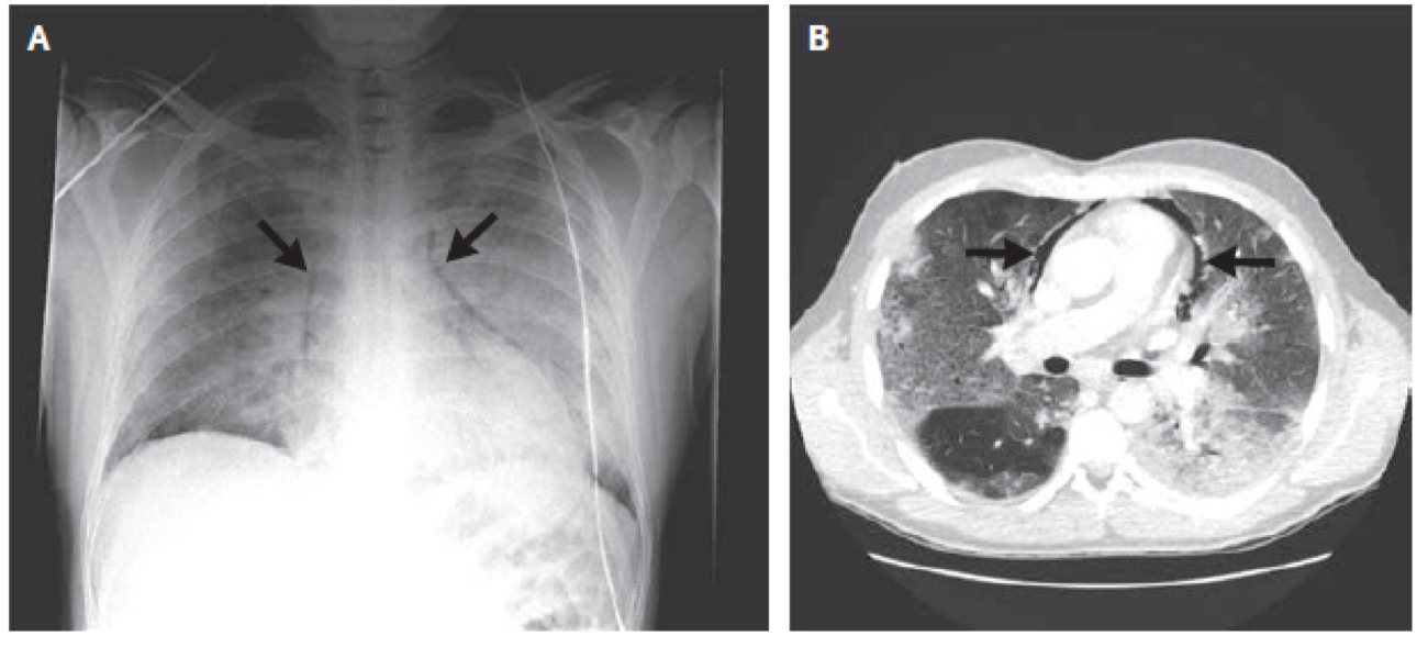
A previously healthy 33-year-old man presented to the emergency department with a 10-day history of lethargy and feeling unwell. His temperature was 38.3°C, and he had tachycardia, with an oxygen saturation of 85% while breathing 15 liters of oxygen per minute through a nonrebreather mask. The physical examination was notable for bronchial breath sounds in the left lower lung; no subcutaneous emphysema was palpated. Chest radiography showed perihilar opacities in both lungs with free air in the mediastinum (Panel A, arrows). Computed tomography confirmed pneumomediastinum (Panel B, arrows) with enhanced interstitial markings and lower-lobe opacities in both lungs, with greater severity in the left lung. The throat swab was positive for influenza A (H1N1). The patient received noninvasive ventilatory support and was treated with zanamivir for influenza and piperacillin–tazobactam for bacterial superinfection. The pneumomediastinum resolved without additional intervention, and the patient was discharged from the hospital 15 days after presentation.
一名既往健康的33岁男性患者因嗜睡及不适10天到急诊就诊。患者体温38.3°C,心动过速,经非重复吸入面罩吸氧15 lpm时氧饱和度85%。体格检查发现左下肺支气管呼吸音;没有发现皮下气肿。胸片显示肺门周围两侧肺组织内阴影伴纵隔游离气体(图A,箭头)。CT证实纵隔气肿(图B,箭头),肺间质纹理增粗,双下肺致密影,左肺为重。咽拭子检查甲型流感病毒H1N1阳性。患者接受无创通气治疗,并使用扎那米韦治疗流感,哌拉西林/他唑巴坦治疗细菌二重感染。纵隔气肿未行干预即消失,15天后患者出院回家。