Cerebral Amyloid Angiopathy
Sihui Chen, Xueping Chen
N Engl J Med 2026;394:1846
DOI: 10.1056/NEJMicm2511044
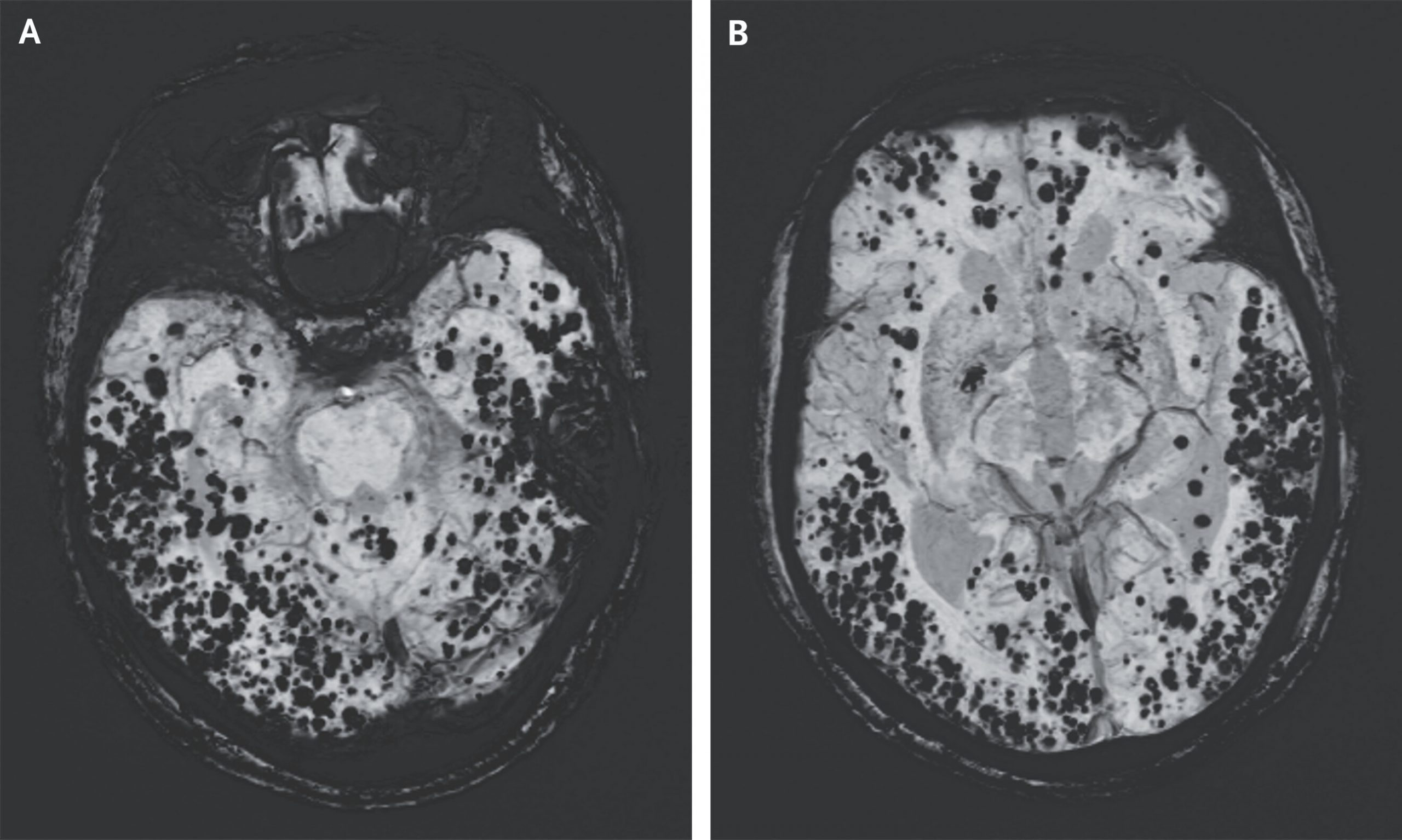
An 86-year-old man presented to the neurology clinic with a 2-year history of progressive memory loss and gait instability. Recent computed tomography of the head had shown mild cerebral atrophy. Neurologic examination was notable for a slow gait with a narrow base of support, as well as moderate-to-severe cognitive impairment on multiple tests. Magnetic resonance imaging with the use of susceptibility-weighted methods showed more than 500 hypointense, round foci throughout the lobar regions, with relative sparing of deep structures, findings consistent with the pattern of cerebral microbleeds seen in cerebral amyloid angiopathy (Panels A and B, susceptibility-weighted imaging sequence). Cerebral amyloid angiopathy is characterized by abnormal deposition of beta-amyloid (Aβ) peptides in the walls of small and medium cerebral blood vessels. Clinical manifestations include acute lobar intracerebral hemorrhage, dementia, and transient focal neurologic episodes. Further diagnostic testing was performed in this patient as part of a clinical trial. Positron-emission tomography showed diffuse cortical amyloid deposition. Analysis of the cerebrospinal fluid showed decreased levels of Aβ42 and Aβ40 proteins and a normal ratio of the two proteins. Exome sequencing was negative for variants associated with cerebral amyloid angiopathy, which is sporadic in most patients. Treatment with supportive care was advised. During the 3 years after diagnosis, the patient had worsening cognitive decline.
