Pneumocystis jirovecii Pneumonia
Ke Xiao
N Engl J Med 2026;394:382
DOI: 10.1056/NEJMicm2514525
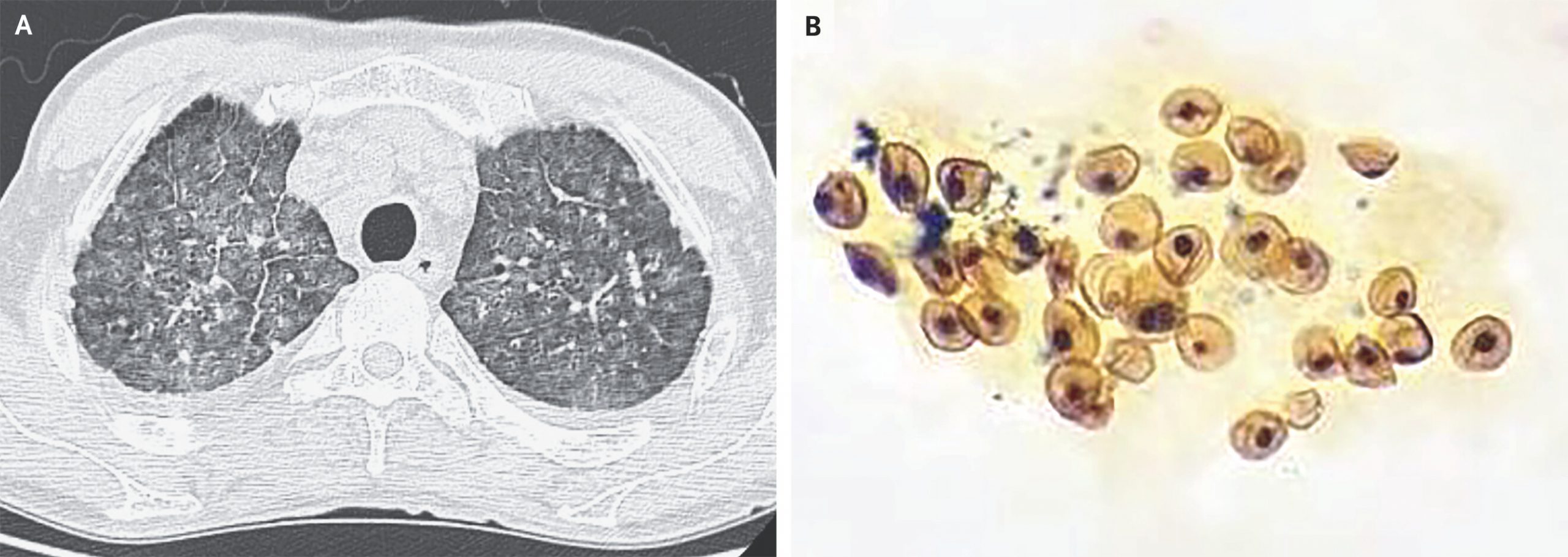
A 60-year-old man with recently diagnosed human immunodeficiency virus (HIV) infection presented to the infectious diseases clinic with a 10-day history of fever, dry cough, and shortness of breath. Treatment for HIV infection had not yet been started. His respiratory rate was 32 breaths per minute, heart rate 120 beats per minute, blood pressure 105/75 mm Hg, and oxygen saturation 89% while he was breathing ambient air. The physical examination revealed cyanosis of the lips but no abnormalities on lung auscultation. A chest radiograph showed diffuse interstitial infiltrates. Computed tomography of the chest showed diffuse nodules and ground-glass opacities with thickened interlobular septa (Panel A). Laboratory studies were notable for a CD4 count of 56 cells per microliter (reference range, 550 to 1440), HIV load of 76,200 copies per milliliter (reference value, <20) and elevated 1,3-β-d-glucan and lactate dehydrogenase levels. Gomori methenamine silver staining of bronchoalveolar lavage fluid showed multiple round structures with thick walls (Panel B), a finding consistent with the cystic form of the fungus Pneumocystis jirovecii. Metagenomic sequencing of the bronchoalveolar lavage fluid confirmed the diagnosis of P. jirovecii pneumonia. Treatment with trimethoprim–sulfamethoxazole and methylprednisolone was started. Nine days after presentation, the patient died of progressive respiratory failure.
