Inhalation Injury from House Fire Smoke
Juan Huang, Cheng Yang
N Engl J Med 2026;394: e39
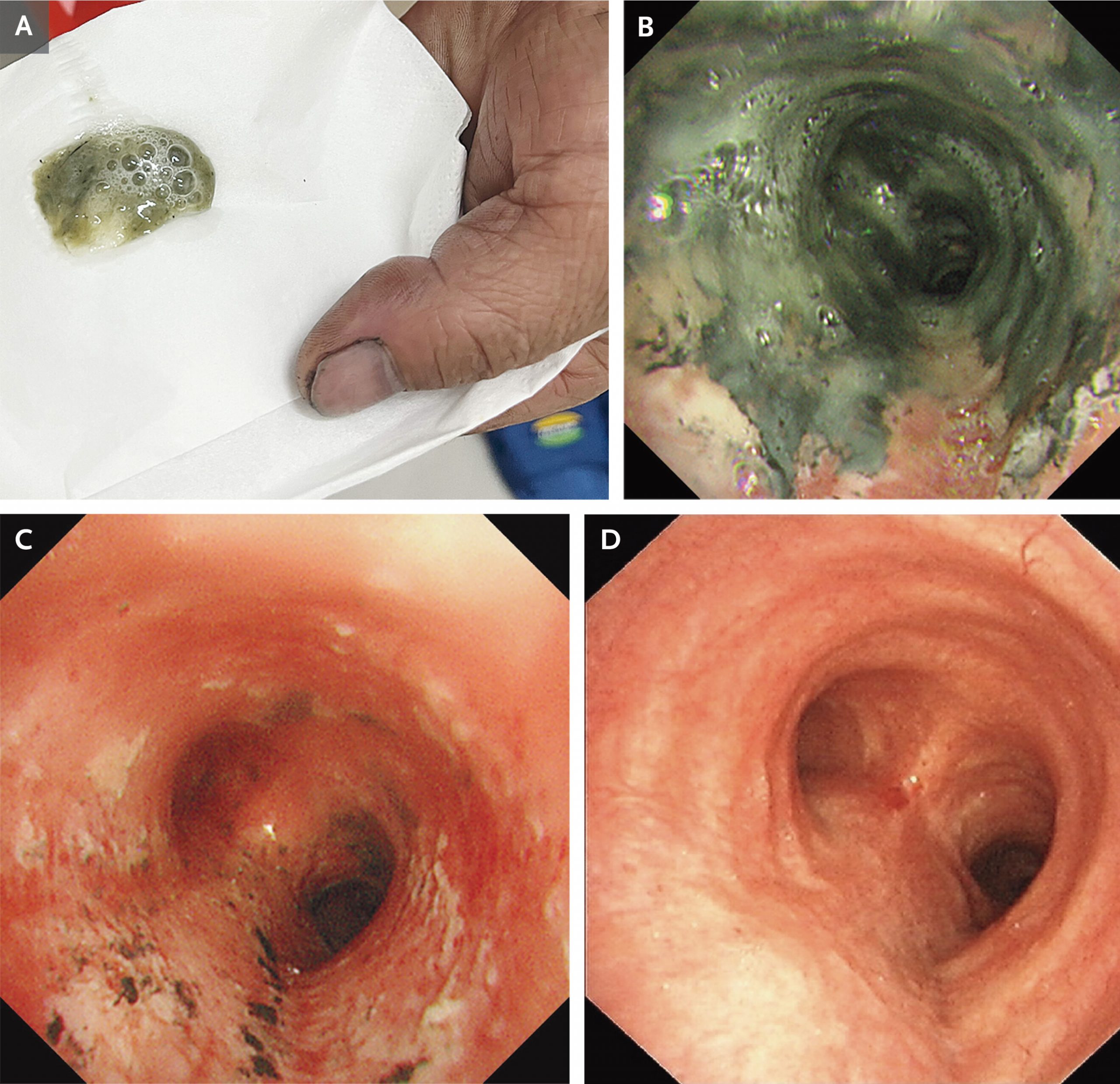
A 55-year-old man presented to the emergency department with cough, chest pain, and worsening dyspnea after being trapped in a burning house for 10 minutes. His heart rate was 120 beats per minute, his respiratory rate 30 breaths per minute, and his oxygen saturation 92% while he was receiving supplemental oxygen through a nasal cannula at a rate of 4 liters per minute. On physical examination, the patient was covered in soot but had no burn injuries. His sputum was carbonaceous (Panel A). Diffuse expiratory wheezing was present on auscultation. Carboxyhemoglobin testing was not available. Computed tomography of the chest revealed segmental mucus plugging. Owing to serious concern about inhalation injury, flexible bronchoscopy was performed to confirm the diagnosis and determine the severity of the injury. After copious secretions were removed from the airway with suction, carbonaceous deposits that extended from the subglottic region to the distal segmental airways were seen (Panel B and video). After therapeutic bronchoalveolar lavage was performed, erythema and edema of the airway mucosa were noted (Panel C). A diagnosis of severe inhalation injury from smoke was made. Treatment with inhaled bronchodilators and supplemental oxygen was administered. The patient was discharged 1 week later. At a follow-up visit 4 weeks after discharge, repeat bronchoscopy showed no abnormalities (Panel D).
