No ventilation, no ARDS: insights from four-dimensional computed tomography as dynamic imaging
Okamura, G., Nishiyama, S., Ono, S. et al.
Intensive Care Med (2024). https://doi.org/10.1007/s00134-024-07643-w
A 63-year-old male, who was in remission from myelodysplastic syndrome after bone marrow transplantation, had previously undergone partial resection of the right upper lobe due to pulmonary aspergillosis. Over a year later, he presented with dyspnea and subsequently required mechanical ventilation for acute respiratory distress syndrome (ARDS) of unknown cause. Neuromuscular blockade was administered, and four-dimensional computed tomography (4D-CT) was performed on day 2.
4D-CT revealed non-ventilated aeration in the right middle lobe, with no ARDS infiltrates observed, contrasting with the other lobes (Fig. 1a–c, supplementary online video 1–2). 4D-CT images and bronchoscopy suggested a bronchial kink of the right middle lobar bronchus, a known post-lobectomy complication (Fig. 1d, supplementary online video 2–3). Although the middle lobe appeared healthy with preserved air volume on conventional CT, 4D-CT as dynamic imaging revealed it to be a non-ventilated lung region.
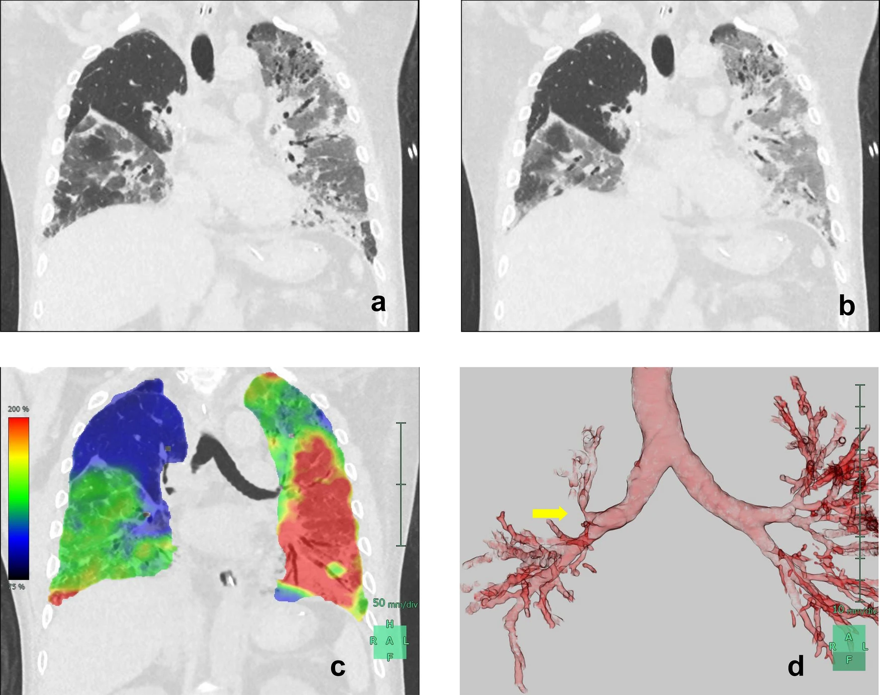
Four-dimensional computed tomography scans showed coronal snapshot images during both inspiratory (a) and expiratory (b) phases. In the right middle lobe, no infiltrative shadows were observed, and aeration was preserved. However, a comparison between inspiratory and expiratory phases revealed that ventilation was not occurring in the right middle lobe. In contrast, in other lung lobes, tidal recruitment occurred due to ventilation. These images were analyzed using software (SYNAPSE VINCENT, Fujifilm, Tokyo, Japan) to construct strain mapping. The degree of alveolar shape distortion during inspiration and expiration was quantified and represents the degree of alveolar strain (c). The index is color-coded with a gradient, displaying smaller values in blue (< 75% of strain) and larger values in red (> 200% of strain). The right middle lobe, which showed low strain, appears in blue. In contrast, the other lung regions are indicated to have high strain. A reconstructed tomographic image showed a stenosis of the right middle lobar bronchus (arrow) (d). These videos are available in the electronic supplementary material for more detailed understanding
This case highlights the importance of identifying non-ventilated areas caused by bronchial kinks and understanding that non-ventilated lung lobes may be spared from conditions typically causing ARDS. Dynamic imaging modalities are useful when optimizing ventilation strategies for ARDS. Although electrical impedance tomography is also used for dynamic imaging, its low spatial resolution hinders detailed anatomical assessment. 4D-CT can provide this crucial information.